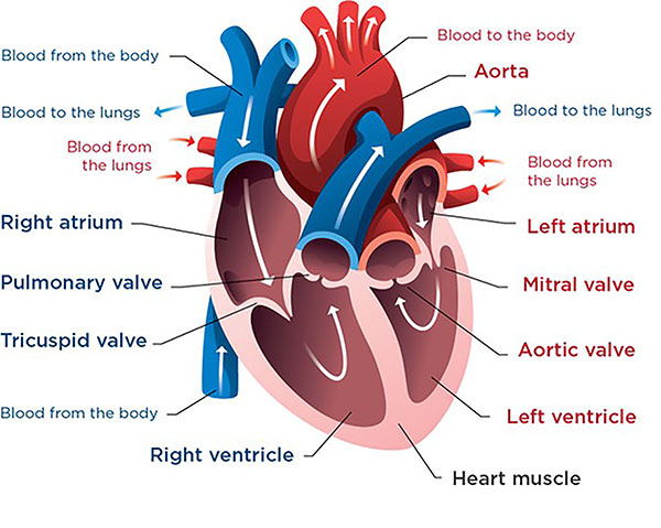
Atrioventricular canal defect is a combination of heart problems resulting in a defect in the center of the heart. The condition occurs when there's a hole between the heart's chambers and problems with the valves that regulate blood flow in the heart.
The large opening in the centre of the heart lets oxygen-rich (red) blood from the heart's left side - blood that's just gone through the lungs - pass into the heart's right side. There, the oxygen-rich blood, along with venous (bluish) blood from the body, is sent back to the lungs. The heart must pump an extra amount of blood and may enlarge. Most babies with an atrioventricular canal don't grow normally and may become undernourished. Because of the large amount of blood flowing to the lungs, high blood pressure may occur there and damage the blood vessels.
Untreated, atrioventricular canal defect can cause heart failure and high blood pressure in the lungs.
Atrioventricular canal defect can involve only the two upper chambers of the heart or all four chambers.
These signs and symptoms are generally similar to those associated with heart failure and might include:
- Fatigue
- Poor weight gain
- Pale skin color
- Irregular or rapid heartbeat
- Excessive sweating
- Difficulty breathing or rapid breathing
Atrioventricular canal defect occurs before birth when a baby's heart is developing. Some factors, such as Down syndrome, might increase the risk of atrioventricular canal defect.
Valves control the flow of blood into and out of the chambers of your heart. These valves open to allow blood to move to the next chamber or to one of the arteries, and close to keep blood from flowing backward.
Atrioventricular canal defect might be detected before birth through ultrasound and special heart imaging.
If your baby is experiencing the signs and symptoms of atrioventricular canal defect, your doctor might recommend:
- Electrocardiogram (ECG or EKG)
- Echocardiogram
- Chest X-ray
- Cardiac catheterization
Surgery is needed to repair complete and partial atrioventricular canal defects. The procedure involves closing the hole in the septum with one or two patches. The patches stay in the heart permanently, becoming part of the septum as the heart's lining grows over them.
For a partial atrioventricular canal defect, surgery also involves repair of the mitral valve, so it will close tightly. If repair isn't possible, the valve might need to be replaced.
For a complete atrioventricular canal defect, surgery also includes separation of the single valve into two valves, on the left and right sides of the repaired septum.
BestHeartSurgery is a comprehensive information portal that gives both the common man and medical professionals.

