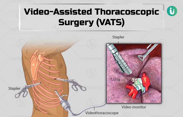
VATS is a surgical procedure used in the chest and lungs. It is a type of 'keyhole' surgery where only very small cuts (incisions) are made to the body. VATS uses a special instrument called a thoracoscope. VATS uses a special instrument called a thoracoscope. This is a thin, tube-like instrument which shines light from the end inserted into the patient. It also transmits images back to an eyepiece or video display so the surgeon can see into the chest cavity. VATS can be used to do a wide range of things, including take small samples of tissues (biopsies) from the lungs. These samples can then be examined in the laboratory.
The surgeon makes two or three small cuts (incisions) in the chest wall near the ribs. These holes are known as ports and are usually about 2 cm long. The surgeon then inserts the thoracoscope through one hole. The thorascope allows the surgeon to see inside the chest. Usually he/she will also insert special surgical instruments into the other incisions. These instruments can be used to remove tissue which may have been seen on an X-ray, or fluid found in the chest. Once the surgery has finished, the instruments are removed and the incisions are closed, usually with stitches.
It is usually easier for patients to recover from video-assisted thoracoscopic surgery (VATS) compared with normal chest surgery (often called 'open' surgery) because the wounds from the cuts (incisions) are much smaller. Air leaks from the lung that don't heal up quickly can keep you in the hospital a longer time and occasionally require additional treatment. A very small number of patients have significant bleeding requiring a transfusion or larger operation.

BestHeartSurgery is a comprehensive information portal that gives both the common man and medical professionals.

